
S2K Commerce - Products Dropdown
Actions
S2K Commerce - Shopping Cart
Actions
S2K Commerce - Order Entry
Actions
Globe Cryo Vials

Globe Cryo Vials
Innovation for the long-term protection of your precious samples.
You've spent days, months, even years getting your labor of love to this point . . . don't risk losing your precious samples to something as simple as a problematic o-ring!
Globe Scientific CryoClear™ vials feature an innovative polyethylene (PE) screw cap that is co-molded with a layer of thermoplastic elastomer (TPE). This special manufacturing process provides the leak-proof benefits required, while eliminating the risk of contamination and sample loss associated with the use of separate o-rings.
Store your samples with the utmost confidence using CryoClear™ cryogenic vials: the best and most leak-resistant cryogenic vials available for the long-term storage of samples at temperatures as low as -196°C.
- Lot certified to be human DNA free, RNase free, DNase free, pyrogen free and ATP free
- Non-pyrogenic, non-cytotoxic, non-mutagenic, heavy-metals free and BSE/TSE free
- Manufactured, assembled and packaged in an ISO 7 (Class 10,000) cleanroom
- Sterilized by beta radiation (SAL 10-6)
- Enhanced cap features a star socket designed specifically for use with automated capping/decapping equipment
- Slim profile cap design ensures better fit in racks and boxes
- Unique barcode printed on each vial for automated data collection
- White writing surface for specimen identification
- Printed graduations for accurate measurements
- Vials and caps are autoclavable
- Produced from medical grade raw materials that will not discolor after re-sterilization
- Tamper evident packaging
- Self-standing tubes interlock with Globe workstation racks

| SKU Number | Vol of Tube | Color | Thread and Bottom | Qty | Price | Quantity | Add to Cart |
| GLB-CRYO-1ML | 1 mL | Natural [Sterile] | External Thread Self Standing | 500 Vials |
287.9000
170.00
Each
|
||
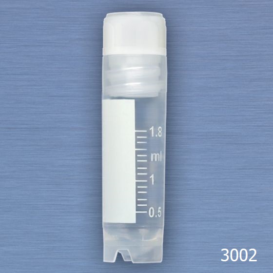
| 3002 | 2 mL | Natural [Sterile] | Internal Thread Self Standing | 500 Vials |
287.90
Each
|
||
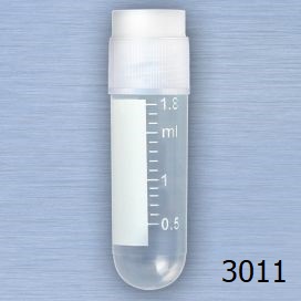
| 3011 | 2 mL | Natural [Sterile] | External Thread Round Bottom | 500 Vials |
287.9000
170.00
Each
|
||
| 3020-2 | 2 mL | Natural [Sterile] | External Thread Self Standing | 500 Vials |
473.30
Each
|
Web Content Viewer
Actions
